Ventricles Of The Brain Ct
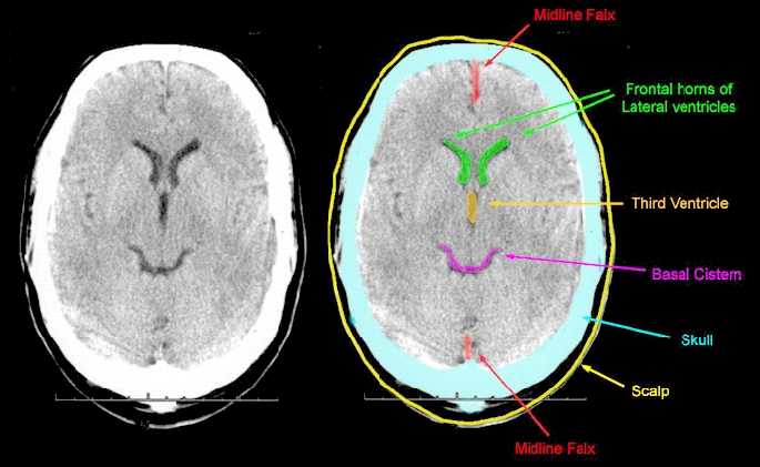
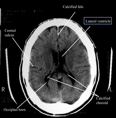
The sulci fissures basal cisterns and ventricles together form the csf spaces also known as the extra axial spaces. Shift the falx should be in the midline with ventricles the same on both sides.
 Native Brain Computed Tomography Small Lateral Ventricles In
There are all together four ventricles in the human brain that constitute the ventricular system along with the cerebral aqueduct.

Ventricles of the brain ct. Central image obtained from wikipedia 4 neuroanatomy. As a follow up to the previous article on the anatomy and physiology of the brain the hydrocephalus association would like to continue our learning and explore the terms we so often hear when dealing with hydrocephalus. It is within the choroid plexus that csf is produced.
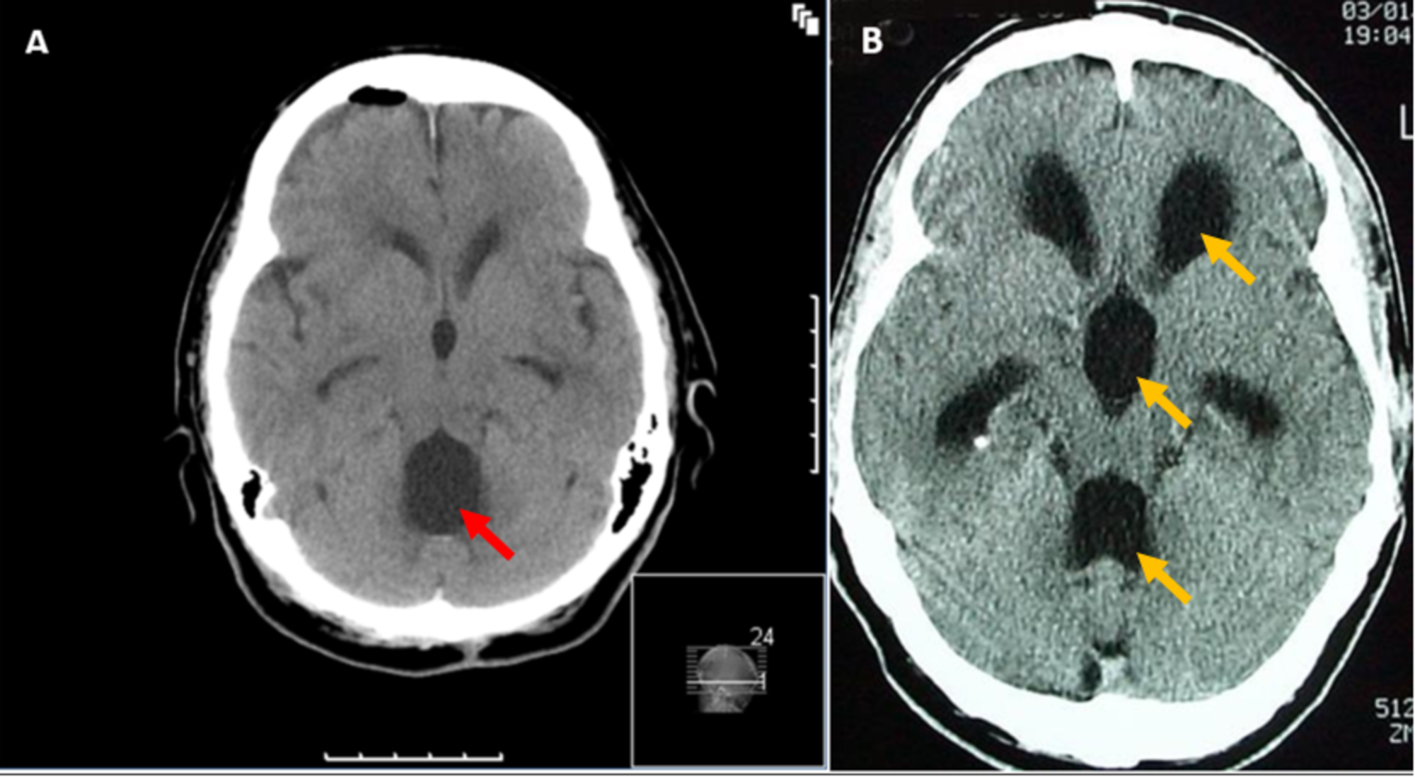
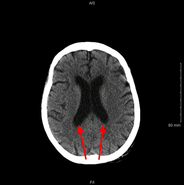
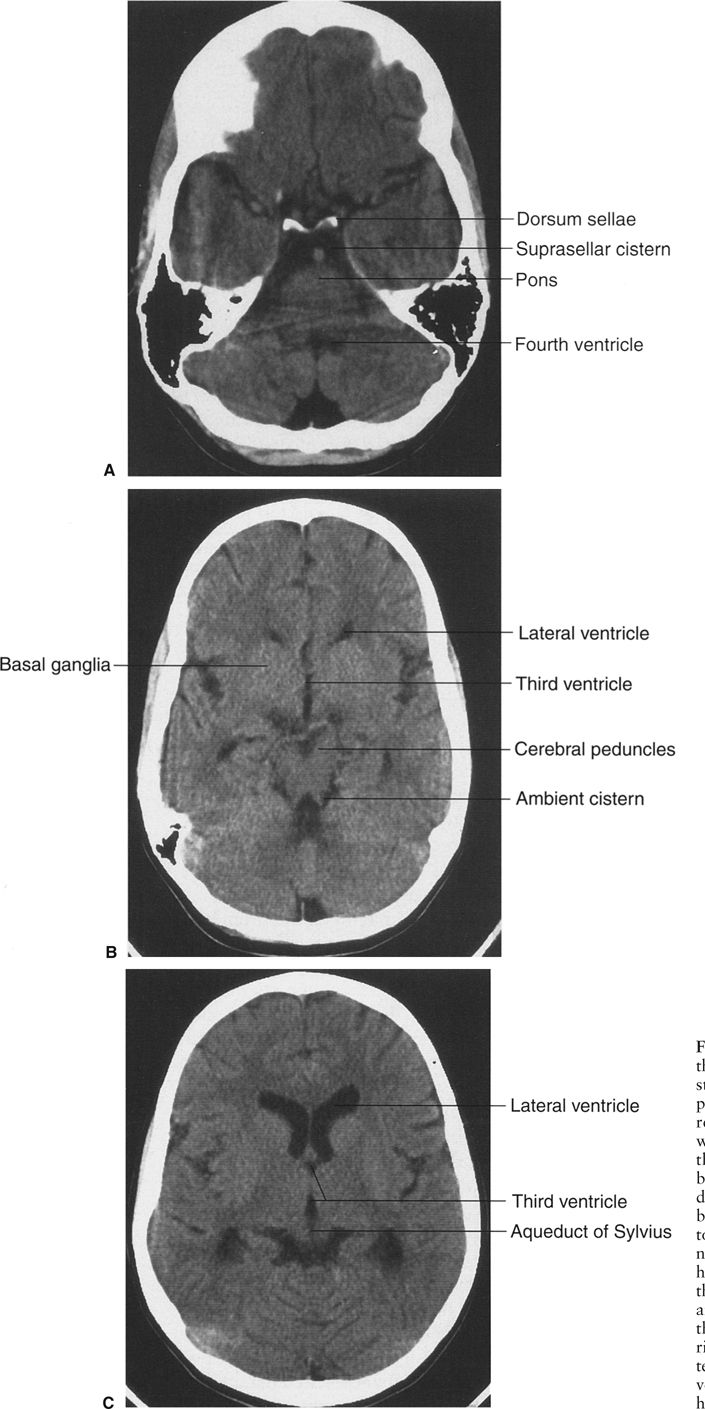
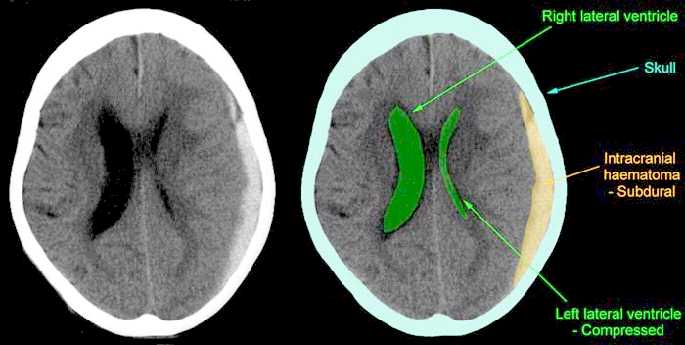
These mass lesions are located in areas that are difficult to reach surgically and because they are intraventricular spread via the csf is common. Compare side to side. Level of the lateral ventricles.
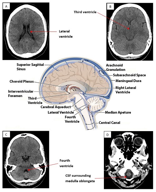
The two largest are the lateral ventricles in the cerebrum the third ventricle is in the diencephalon of the forebrain between the right and left thalamus and the fourth ventricle is located at the back of the pons and upper half of the medulla oblongataof the hindbrain. The ventricles of the brain the ventricles are structures that produce cerebrospinal fluid and transport it around the cranial cavity. They are lined by ependymal cells which form a structure called the choroid plexus.
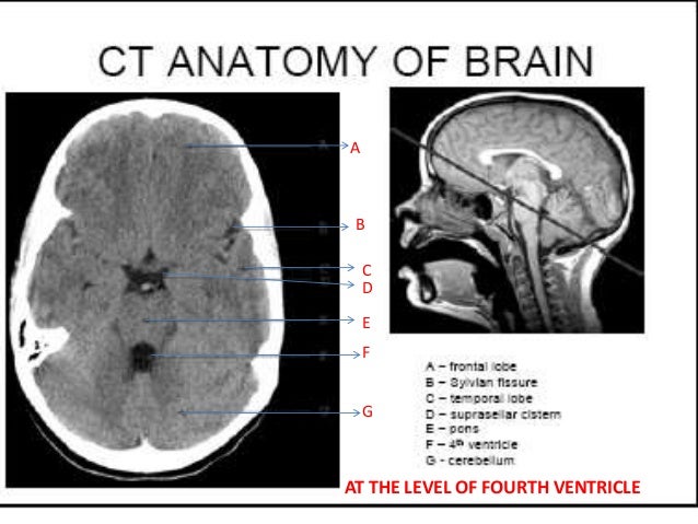
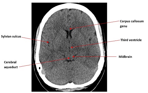
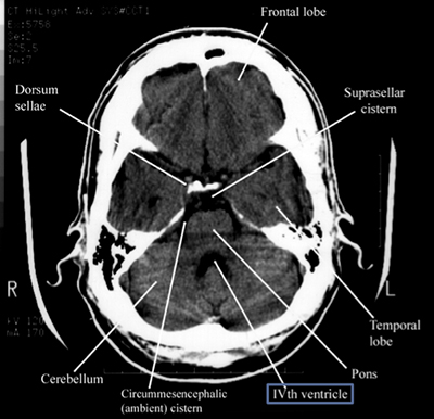
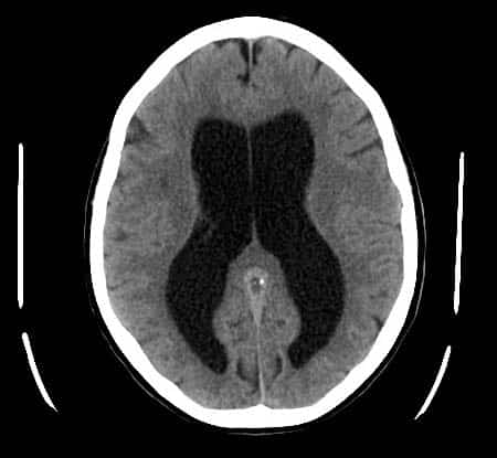
The below ct axial slices highlight common anatomical structures which are helpful to know when interpreting a ct brain scan. Level of the spinal cord. An appreciation of the normal appearances of the csf spaces is required to allow assessment of brain volume.
The four cavities of the human brain are called ventricles. The ventricles and csf flow. Level of the fourth ventricle.
Grey white differentiation the earliest sign of a cva on ct scan is the loss of the grey white interface on ct scan. Ct and mr imaging have been useful in demonstrating these masses but imaging characteristics are usually nonspecific. Check for effacement of sulci unilateral or bilateral.
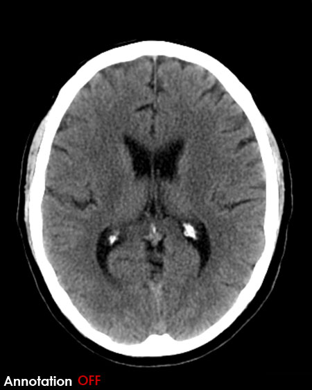
Ventricles are hollow cavities of the brain that contain the cerebrospinal fluid csf which circulates within the brain and spinal cord. Csf is of lower density than the grey or white matter of the brain and therefore appears darker on ct images. Ct brain images ct appearances of the cerebral ventricles including the lateral ventricles third ventricle fourth ventricle basal cisterns and cisterna magna.
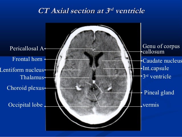
One tenth of all cns neoplasms involve the ventricles of the brain. Level of third ventricle.
 Cureus Disproportionately Large Communicating Fourth Ventricle
 A Morphometric Study Of Ventricular System Of Human Brain By
 Memory Loss With Enlarged Brain Ventricles Bmj Case Reports
 Brain Imaging
 Normal Ct Brain
 Radiological Anatomy Techniques Of The Ventricular System
 Ct Brain Scroll Image Gallery Normal Ventricles
 A Morphometric Study Of Ventricular System Of Human Brain By
 How To Read A Head Ct Emergency Medicine Newyork Presbyterian
 Ventricular System Radiology Reference Article Radiopaedia Org
The Ventricular System Of The Brain Radiologypics Com
 Subependymoma Lateral Ventricle Radiology Case Radiopaedia Org
 Brain Imaging
 Trigone Of Lateral Ventricle Normal Brain Ct Radiology Gallery
 Pdf Measurement Of Normal Brain Lateral Ventricles Byusing Ct
 The Ventricles Neuroanatomy Video Lab Brain Dissections Youtube
 A Morphometric Study Of Ventricular System Of Human Brain By


 Ct Axial Image Of The Brain Showing The Length Of Body Of The
 How To Read A Head Ct Emergency Medicine Newyork Presbyterian
 Hydrocephalus Simply Radiology

 Brain Radiology Key
 Measurements Of Cerebral Ventricle Indices A Maximum Bifrontal
 Lateral Ventricle Radiology Reference Article Radiopaedia Org

 Brain Ventricle Parcellation Using A Deep Neural Network
Https Encrypted Tbn0 Gstatic Com Images Q Tbn 3aand9gctvvoq914ybhnavfnbtps4rcay900y2hdc4bmdt4sajvpqbyv P Usqp Cau
 Ct Axial Image Of The Brain Showing The Length Of Body Of The
 Memory Loss With Enlarged Brain Ventricles Bmj Case Reports

 Normal Anatomy Radiology Key
 The Ventricles Of The Brain Lateral Third Fourth
Posting Komentar
Posting Komentar